OrthoCarolina and Tryon Medical Partners are named “Official Healthcare Providers” of the Carolina Ascent FC
OrthoCarolina and Tryon Medical Partners are proud to serve as the Official Healthcare Providers of Carolina Ascent FC, delivering trusted, comprehensive care to support athlete health, performance, and community engagement.
Read More
OrthoCarolina Launches Premier Orthopedic Physician Assistant Fellowship
A first-of-its-kind fellowship provides immersive clinical and surgical training under nationally recognized orthopedic specialists, elevating PA training and patient care
Read More
OrthoCarolina Partners with Yes I Can Basketball to Provide Scholarships and Keep Youth Sports Accessible in Charlotte
OrthoCarolina has partnered with Yes I Can Basketball to provide scholarships and ensure that youth sports in Charlotte remain accessible. This collaboration supports athlete development, injury prevention, and community well-being through affordable programs and resources.
Read More
Introducing PT Solutions – the New Preferred Therapy Provider of OrthoCarolina
We are excited to announce that OrthoCarolina has selected a new preferred provider of physical and occupational therapy services. Beginning February 3, 2025, PT Solutions will provide you with the same exceptional care you’ve become accustomed to at OrthoCarolina.
Read More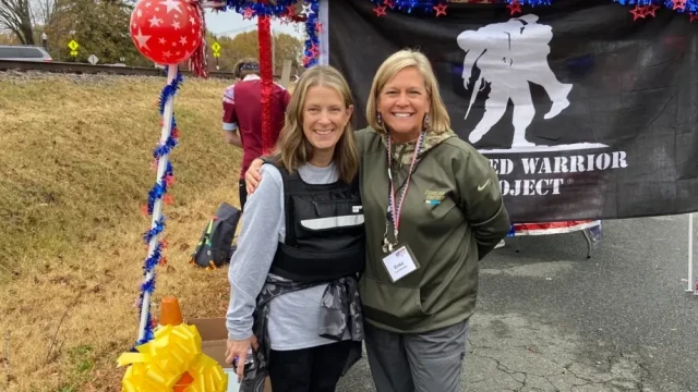
Championing Veterans' Health: The Impact of OrthoCarolina's Partnership with the Wounded Warrior Project
OrthoCarolina partners with the Wounded Warrior Project to support veterans' physical and mental health. Learn how their orthopedic care, advocacy, and community efforts are transforming lives and providing essential resources for those who have served.
Read More
OrthoCarolina Shines with 36 Physicians Named Charlotte's "Top Doctors" for 2024!
Each year, Charlotte Magazine publishes its highly anticipated "Top Doctors in Charlotte" list, honoring exceptional medical professionals based on a respected peer survey by Castle Connolly. For 2024, 36 OrthoCarolina surgeons have been recognized for their expertise and dedication, with many earning repeat honors that underscore their ongoing commitment to excellence. Join us in celebrating these outstanding OrthoCarolina doctors!
Read More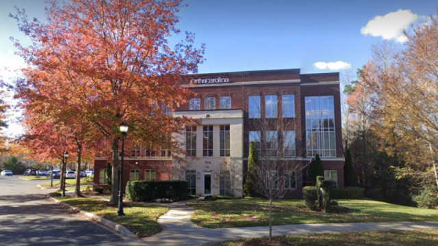
OrthoCarolina Expands Regional Reach, Enhances Patient Value with Acquisition of Matthews Surgery Center
OrthoCarolina, a leading orthopedic practice, announced its acquisition of Matthews Surgery Center from Novant Health, effective October 1, 2024. The specialized facility offers advanced orthopedic surgery and pain management for adults and children, featuring state-of-the-art technology and expert care.
Read More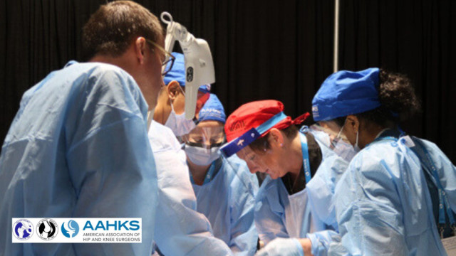
Elevating Excellence: The Adult Reconstruction Fellowship Recognition Program

How OrthoCarolina Supports Pit Crews Weekly for Peak Performance
Discover how OrthoCarolina supports motorsports pit crews with on-site care, injury prevention, and tailored health plans to keep elite athletes in top shape for every race.
Read More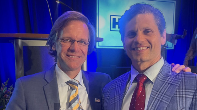
World-Renowned OrthoCarolina Physicians Create First-of-its-Kind Multidisciplinary Multi-Extremity Limb Loss Program
The program uses a comprehensive approach including proprietary surgical techniques and virtual reality therapy to rapidly restore function and reduce residual and phantom limb pain.
Read More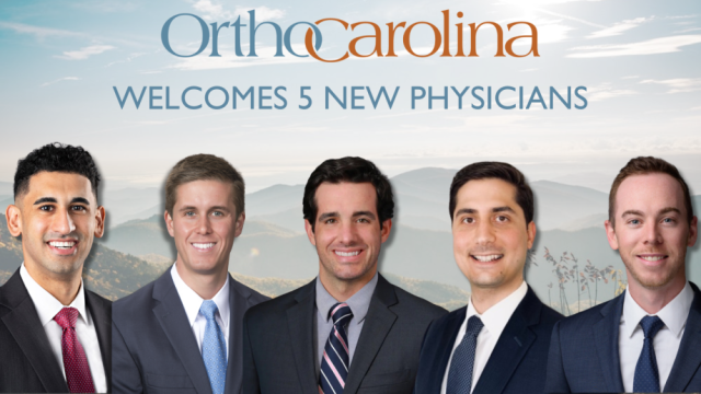
OrthoCarolina Welcomes Five New Doctors
OrthoCarolina is excited to announce the addition of five expert physicians to our team starting September 1st, 2024. Discover more about Dr. Gregory Pereira, Dr. Justin Rennard, Dr. Petros Koutsogiannis, Dr. Ryan McCarter, and Dr. J. Taylor Bellamy, and the exceptional care they will bring to our communities.
Read More
Strengthening Motorsports with OrthoCarolina and Spire Motorsports
Discover how the partnership between OrthoCarolina Motorsports and Spire Motorsports is revolutionizing health and wellness in NASCAR. Learn about the specialized orthopedic care provided by OrthoCarolina's experienced team, ensuring the physical well-being of drivers, pit crews, and their families. Explore how this collaboration is driving peak performance, minimizing injuries, and setting a new standard in motorspor
Read More
OCRI's Impact: Celebrating National Nonprofit Day with OCRI and OrthoCarolina
On August 17, National Nonprofit Day, OCRI proudly highlights its diverse range of programs dedicated to making a positive impact in the community. From charitable initiatives and volunteer programs to community outreach and development projects, OCRI's commitment to social good and philanthropy is showcased through its numerous achievements and success stories. Join us in celebrating and supporting the efforts of OCRI as we continue to drive meaningful change and development in our communities.
Read More
From Classroom to Court: Essential Tips to Get Kids Back to School and Back to Sports
Discover orthopedic tips from OrthoCarolina to prepare your child for the upcoming school sports season. Learn how to prevent injuries and enhance performance with expert advice on sports physicals, conditioning, core strength, proper technique, and more.
Read More
OrthoCarolina Belmont Relocates to Mount Holly - New Facility Opening August 2024
OrthoCarolina Belmont is relocating to Mount Holly! Join us at our new facility starting August 26, 2024, for the same exceptional orthopedic care and personalized attention.
Read More
Get Ready for 2024 High School Football: Charlotte Kickoff Night Powered by OrthoCarolina Kicks Off August 23
Join us on August 23, 2024, for the 11th Annual Charlotte Kickoff Night at Memorial Stadium. Watch Hough vs. Northwestern at 5:15 PM and Ardrey Kell vs. Providence at 8:00 PM. Sponsored by OrthoCarolina, providing top medical support for our athletes.
Read More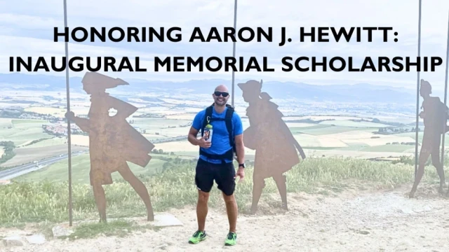
Honoring Aaron J. Hewitt: Inaugural Memorial Scholarship
Discover the legacy of Aaron J. Hewitt with the inaugural memorial scholarship awarded to Scot Rheinecker, PA-C at OrthoCarolina. Celebrate excellence, leadership, and compassion within our community.
Read More
OrthoCarolina Receives The Triple Crown in Research Awards: Advancing Orthopedic Care Through Groundbreaking Research
Explore OrthoCarolina's groundbreaking research, winning top awards from AAOS and AAHKS. Discover how our innovative studies are transforming orthopedic care, from revolutionizing treatment protocols for prosthetic joint infections to advancing opioid-free pain management in spine surgery.
Read More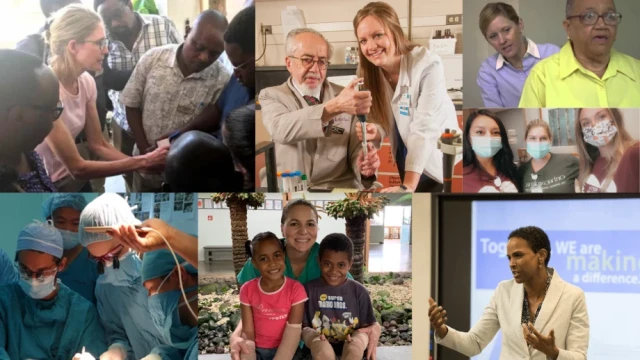
Celebrating Women Physicians at OrthoCarolina Through Women Physician Month
Explore the groundbreaking contributions of Women Physicians at OrthoCarolina throughout February. Celebrating diversity in the orthopedic field, discover the expertise and dedication of these trailblazing specialists, shaping a future of medical empowerment.
Read More
50 OrthoCarolina Doctors Named Top Docs!
Discover the 50 distinguished OrthoCarolina Physicians who have achieved the esteemed 'Top Docs' recognition in Charlotte. These dedicated medical professionals have been honored based on a peer survey conducted by Castle Connolly and are at the forefront of healthcare excellence in the region
Read More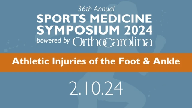
OC Presents the 2024 Sports Medicine Symposium
The 36th Annual Sports Medicine Symposium, presented by OrthoCarolina, will be held on Saturday, February 10, 2024, at the Dale F. Halton Theater.
Read More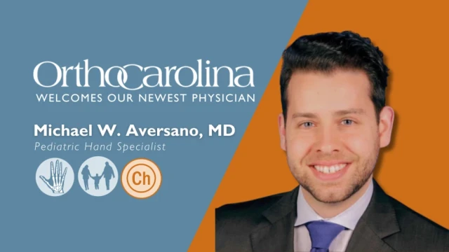
Welcome Dr. Aversano!
Discover the expertise of Michael W. Aversano, MD, a distinguished Pediatric Hand Specialist at OrthoCarolina. Explore his extensive background, publications, presentations, and contributions to the field of orthopedic surgery.
Read More
OrthoCarolina Excels in Charlotte's Best 2023: When better is BEST... Charlotte's Best!
OrthoCarolina shines in Charlotte's Best 2023 awards, claiming Gold in Orthopedics. Discover our award-winning healthcare services and career opportunities in Charlotte and the Carolinas.
Read More
OrthoCarolina Appoints Leo Spector, MD, MBA as Next CEO
Discover the latest leadership transition at OrthoCarolina as Dr. Leo Spector, a distinguished spine surgeon and visionary in Value-Based Care, assumes the role of CEO, succeeding Dr. Bruce Cohen.
Read More